Abdominal/Pelvic CT Scans

Abdominal/Pelvic CT Scans
CT scanning has drastically changed the evaluation of abdominal disorders and has decreased dramatically the number of unnecessary laparotomies, both traumatic and nontraumatic. It has allowed us to open the “black box” which hides many catastrophic processes, guiding or changing management. While a complete discussion of use of abdominal trauma CT is beyond the scope of this chapter, selected emergency conditions will be presented.

Overview
Patient Preparation

  • Patients undergoing abdominal CT for either traumatic or nontraumatic conditions should ideally have oral contrast prep with adequate bowel transit time to allow for visualization of the bowel. The oral contrast is not necessary for investigation of renal colic or abdominal aneurysm, and is optional for traumatic conditions if time does not permit.
  • Intravenous contrast is usually given for abdominal CT, but is not necessary for the majority of renal stone CTs, unless more detail of the ureteral anatomy is needed.
    Intravenous contrast should be avoided in the setting of renal insufficiency for fear of renal contrast damage or in the presence of the oral hypoglycemic metformin for fear of lactic acidosis.
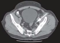
Distal Left Ureter Stone

Figure: Distal left ureter stone (white arrow).
Renal Colic

Since the advent of helical CT, it has quickly replaced intravenous pyelography (IVP) as the initial investigation of suspected renal colic. It can be performed quickly, without the need for patient preparation, or laboratory investigations such as BUN/ creatinine and costs less than IVP.

  • Findings of renal calculus on unenhanced abdominal CT:
  • Renal calculus visualized.
  • Hydronephrosis will be seen with an obstructing ureteral stone.
  • Perinephric stranding or fluid may be seen with increasing grades of obstruction.
  • Advantages of helical CT:
  • Fast scanning time.
  • No intravenous contrast exposure.
  • No need for patient preparation such as precontrast labs or bowel prep.
  • May identify alternative diagnoses, changing management such as abdominal aneurysm, appendicitis, and diverticulitis.
  • Cost is lower in most institutions than IVP.
  • Disadvantages of helical CT include:
  • Radiation exposure is greater than with IVP.
  • May not identify small nonobstructing stones.
  • May not identify some noncalcified stones such as uric acid stones, medication-related stones such as indinivir. Does not show renal function as with IVP.
  • Alternative imaging:
  • Ultrasound
  • Intravenous pyelography

    Left Hydronephrosis


    Figure: Left hydronephrosis seen on CT scan due to obstructing ureter \stone.
    Note the enlarged renal pelvis compared with the right kidney.

Acute Appendicitis
Appendicitis is an excellent example of how imaging has changed the practice of medicine. While it has been acceptable to have negative laparotomy rates of up to 30%, this number has come down substantially in those institutions utilizing CT scanning in the investigation of right lower abdominal pain. While initial studies were performed on selected populations, adding selection bias, high sensitivities and specificities have seemed to hold up in the general population. Like its use in renal colic, it may also reveal alternate diagnoses, thereby obviating exploratory laparotomy/laparoscopy.
Appendiceal CT scanning is performed using helical technique with thin slices through the region of the appendix after the patient has received adequate oral contrast. Rectal contrast is often used to ensure filling of the cecum. Intravenous contrast is commonly used but may be omitted if contraindicated.

  • Signs of appendicitis on CT scan include:
  • Thickened appendiceal wall greater than 2 mm.
  • Distended appendix measuring greater than 6 mm.
  • Appendix not filling with contrast despite adequate terminal ileal and cecal visualization.
  • Inflammatory changes surrounding the appendix are suggestive, but not diagnostic, of appendicitis as this may occur with nonsurgical conditions.
  • Fluid collection or abscess in the right lower quadrant is also suggestive of appendicitis.
  • As with any other test, it is important to understand its limitations and not rule out appendicitis solely on a negative CT, in the face of high clinical suspicion. Possible reasons for a false negative CT scan include:
  • Inadequate patient preparation not allowing for visualization or filling of the appendix.
  • Thin patient not providing enough fat stranding to appreciate inflammatory changes.
  • CT scan performed too early!! Not enough time may have lapsed to allow the inflammatory changes to be seen on CT.
  • As with any imaging study, its power lies in those interpreting the scan. Reading appendiceal CT requires skill and experience.
  • Advantages of CT for appendicitis:
  • May provide alternate, nonsurgical diagnosis thereby obviating surgery.
  • Noninvasive diagnosis, unlike laparoscopy.
  • May be more cost-effective than in-hospital observation or surgical exploration.
  • Disadvantages of CT scan in appendicitis:
  • Radiation exposure is significant, averaging around 3- 5 rems.
  • May delay treatment in those with obvious clinical diagnosis if ordered inappropriately.

    Acute Appendicitis


    Figure: Acute appendicitis. The nonfilling appendix is seen entering the cecum.

    Acute Appendicitis Associated Inflammatory Fat Stranding


    Figure: Acute appendicitis. This cut shows the appendix with associated inflammatory fat stranding.

  • Alternate imaging studies in appendicitis:
  • Ultrasound
  • Laparoscopy

Abdominal Aneurysm

  • Although treatment decisions regarding abdominal aneurysm repair are often made using clinical symptoms and signs, CT scanning can be an appropriate imaging study.
  • CT scanning is very useful for evaluating suspected or known abdominal aneurysm patients without indications for immediate operative management. It provides detail of the aneurysm dimensions and location, and can often reliably comment on leaking or rupture.
  • In the setting of operative indications, especially with a known aneurysm, obtaining a CT scan will only delay definitive surgical treatment.
  • Although an abdominal aneurysm will be identified without the aid of intravenous contrast, details such as leaking, rupture, thrombosis or dissection may not be identified.
  • Findings of abdominal aneurysm on CT scan:
  • Abdominal aorta measuring greater than 2.5 cm in diameter or greater than 1.5 times the diameter of adjacent aorta.
  • Calcifications are common in the lumen due to associated atherosclerotic disease.
  • Associated thrombus may be seen lining the lumen of the aneurysm.
  • An intimal flap may be seen with associated dissection.
  • Retroperitoneal or free peritoneal leak may be seen.

    Large Abdominal Aneurysm


    Figure: Large abdominal aneurysm is identified on this noncontrast CT scan of the abdomen

  • The location and extent of aneurysm (i.e., involvement of renal arteries) as well as associated leak are important in surgical planning and should be conveyed to the consultant.
  • Advantages of CT scan in abdominal aneurysm evaluation:
  • High sensitivity.
  • Able to provide dimensions and extent of aneurysm.
  • Unlike ultrasound, can often identify complications such as leak, rupture or dissection.
  • Disadvantages of CT scan in abdominal aneurysm evaluation:
  • Usually requires patient to be transported out of the department.
  • Since it provides helpful details, consultant surgeons may try to “push” for a CT before definitive treatment.
  • Pitfalls in abdominal CT scan for abdominal aneurysm:
  • Ordering CT scan inappropriately for patient with clear operative indications.
  • Sending a potentially unstable patient to Chest CT scan when a bedside ultrasound would identify the aneurysm.
  • Alternative imaging studies:
  • Bedside ultrasound
  • MRI
  • Angiogram

Small Bowel Obstruction

  • The majority of small bowel obstructions can be diagnosed on plain radiographs and occur in the setting of clear risk factors such as previous surgery or incarcerated hernia.
  • When small bowel obstruction is suspected and is either not clear on abdominal radiographs or occurs without a discernable etiology, CT scanning can provide the necessary details.
  • CT scanning with bowel contrast preparation is about 90% sensitive and specific for bowel obstruction and may reveal uncommon etiologies such as internal incarcerated hernias, intussusception, or volvulus.

Undifferentiated Elderly Abdominal Pain

  • As previously discussed, abdominal CT scanning has allowed clinicians to open the “black box” of the abdomen without need for surgical exploration. One clinical presentation in which this is most evident is undifferentiated abdominal pain, especially in the elderly.
  • The elderly are notoriously difficult to evaluate by conventional means as they often lack both symptoms and clinical and laboratory signs found in serious disease.
  • Despite this, the elderly are much more likely to have a surgical condition causing abdominal pain.

    Loops of Distended Small Bowel


    Figure: Loops of distended small bowel are seen on this CT scan of a small bowel obstruction.

  • Plain abdominal radiographs are often not helpful in young healthy patients with abdominal pain. The elderly, however, often reveal silent abnormalities that may change management, and ordering of plain X-rays should be liberal in this population.
  • CT scan is able to identify many of the surgical conditions causing elderly abdominal pain and is often helpful when conventional investigation and consultation has not produced a diagnosis.
  • A caveat is needed, however, in order that the CT not be used to “rule out” disease in the setting of a “normal” plain abdominal film. It has limitations, as any study, and the reader is cautioned to use the CT scan as one more piece of information and clinical judgement above all.
       
eXTReMe Tracker