|
Summary
- As the speed of image acquisition improves and nonmagnetic critical care monitoring equipment becomes available, MRI will become an increasingly important diagnostic tool of the emergency physician.
- Currently there are several emergent indications for MRI where there is no appropriate substitute, and other indications to consider MRI on a semiemergent basis, and finally indications to substitute MRI for angiography and CT because of contrast contraindications.
- Emergent indications:
- Spinal cord compression—gold standard for nontraumatic cord compression; time-delays in diagnosis may result in irreversible damage.
- Traumatic spinal cord injury—MRI scan is the recommended modality for trauma patients with neurologic symptoms and normal plain films.
- Aortic dissection—One of four possible studies which can be used emergently to diagnose dissection; considered the best all-around study as long as the patient is not unstable.
- Semiemergent indications:
- Dural venous sinus thrombosis—a rare cause of stroke but one which affects primarily young people and has disastrous consequences; although CT scan is the emergent test in stroke patients to exclude bleed, MRI scan, VQ scan should be done soon after if suspicious for DVST because anticoagulation and antibiotics may improve outcome.
- Cervical artery dissections—another rare cause of stroke seen in younger patients;
- MRI/MRA is now the recommended first line study although surgery is rarely performed and anticoagulation is of unclear benefit.
- Spinal cord infections—although antibiotics are typically started based on clinical findings, information gathered from MRI can elucidate whether emergent surgery is necessary (for expanding abscess) to avoid imminent cord compression.
- Encephalitis/vasculitis—a diagnosis of Herpes encephalitis or cerebral vasculitis can be missed and appropriate treatment not started if MRI is not performed so consider in patients with clinical clues for these processes.
- Substitute for angiography:
- Subarachnoid hemorrhage—although angiography is the gold standard for aneurysm location and potential embolization, patients who have contraindications to angiography may undergo MRA as a highly accurate alternative.
Complications
- 5% of the population experience claustrophobia in the MR scanner which can be reduced by mild sedation.
- Unlike CT, movement of the patient during MRI scan will distort all images so patient cooperation is very important.
- The only serious complication of MRI scan is from the interaction of the high magnetic fields with internal ferromagnetic objects (aneurysm clips, pacemakers, etc) or external metal objects which can be attracted to the magnet and act as missiles if brought into the room.
- Gadolinium, the contrast agent used in many MRI studies of the CNS, does not cause renal failure and allergic reactions are extremely rare.
- Common contraindications for MRI
- Cardiac pacemaker or permanent pacemaker leads
- Internal defibrillatory devices
- Cochlear prostheses
- Bone growth stimulators
- Implanted spinal cord stimulators
- Electronic infusion devices
- Intracranial aneurysm clips (some)
- Ocular implants (some) or ocular metallic foreign body
- Omniphase penile implant
- Swan-Ganz catheter
- Magnetic stoma plugs
- Magnetic dental implants
- Magnetic sphincters
- Ferromagnetic IVC filters, coils, stents—considered safe at 6 wk after implant.
- Tattooed eyeliner
Indications for Emergent MRI
Spinal Cord Compression
- The most compelling reason to order an MRI from the ED is for suspected spinal cord compression.
- MRI scan has replaced CT scan myelography as the imaging study of choice for evaluation of the spinal canal because of superior contrast resolution, lack of ionizing radiation, lack
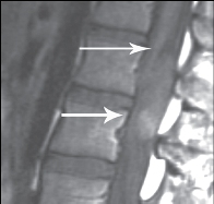
 Figure: Conus ependymoma.
Ependymoma in lower lumbar spinal canal with anterior displacement of the conus medullaris.
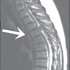
 Figure: Thoracic meningioma
compressing the cord with metastasis noted posterior to the vertebrae (arrow).
of contrast material risk, absence of need for spinal needle placement and ability to detect multiple lesions with minimal patient manipulation.
Cervical Spine Trauma
- The American College of Radiology (ACR) published appropriateness criteria for ordering various imaging tests in cervical spine trauma.
- Their only indication for MRI is a patient with neurological signs and symptoms whose plain films are normal.
Dural Venous Sinus Thrombosis
- DVST is a thrombus formation within any of the major dural venous sinuses that drain blood from the brain.
- DVST most commonly affects young and middle-aged individuals (accounts for 1-2% of strokes in patients under 45) and may occur spontaneously or in association with risk factors.
- MRI and MR venography (a noninvasive technique using contrast) has replaced angiography as the study of choice for the evaluation of suspected DVST.
|
|