|
Anatomy
- The knee is more complex than a simple hinge joint.
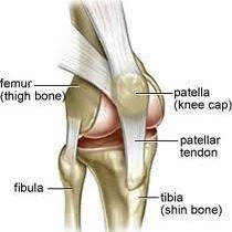
- It is made of three articulations, the patellofemoral, the tibiofemoral, and the tibiofibular joints.
- The ligamentous components of the joint are the lateral collateral ligament (LCL), medial collateral ligament (MCL), the anterior cruciate ligament (ACL), and the posterior cruciate ligament (PCL).
- The extensor mechanism includes the quadriceps tendon, the patella, and the patellar ligament.
- The important cartilaginous structures are the medial and lateral menisci.
- The medial meniscus is connected with the MCL; the lateral menicus is not.
Management
Prehospital
- Prehospital care entails immobilization to prevent further neurovascular injury, elevation and ice.
History
- The emphasis is on mechanism.
- The following are classical injuries associated with common mechanisms of injury:

- Head on traffic collision
|
Posterior cruciate ligament injury |
- Twisting injury (i.e., skier)
|
Anterior cruciate ligament tear |
- Contact with lateral force
(i.e., football player) |
MCL tear, medial meniscus tear and ACL tear
(aka O�Donaghue�s triad) |
- Hyperextension
|
ACL injury followed by PCL injury |
- Turn with tibia rotated
in opposite direction |
Patellar dislocation |
Physical Exam
Radiography
- Plain Films
- The Ottawa Knee Rules were described in order to delineate which patients require plain films.
- Standard views are the AP and lateral.
- Other views may be helpful to elucidate individual injuries if suspected:
- Cross-table lateral
|
May detect fat-fluid level, pathognomonic of fracture |
- Oblique views
|
Fracture or loose foreign body |
- Notch view
|
Osteochondral fracture |
- Sunrise view
|
Patellar injuries |
- Plateau view
|
Tibial plateau fracture |
- CT and MRI are rarely used in emergency imaging of the knee.
- MRI provides superior visualization of soft tissue structures including menisci and cruciate ligaments, but is usually performed by the primary care or orthopedist in a nonurgent setting.

A knee X-ray series is only required for knee injury patients with any of these findings:
- age 55 yr or older, or
- isolated tenderness of patella*, or
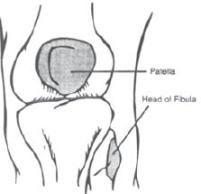
- tenderness at head of fibula, or
- inability to flex to 90�, or
- inability to bear weight both immediately and in the emergency department (4 steps).**
* No bone tenderness of knee other than patella.
** Unable to transfer weight twice onto each lower limb regardless of limping.
Figure 8.4. Ottawa Knee Rule for use of radiography in acute knee injuries. From Stiel et al. Implementation of the Ottawa Knee Rule for the use of radiography in acute knee injuries. JAMA 1997; 278:2075-2079.
Table Classification of ligament sprains
| STRETCH-GRADE I |
A first-degree sprain is really a microscopic tear and can be treated with rest, ice, and
protection with crutches and/or a splint. |
| PARTIAL-GRADE II |
A second-degree sprain should be immobilized to prevent the ligament from tearing
completely. |
| COMPLETE TEAR-GRADE III |
| Third-degree sprains may require surgery. Therapy is somewhat controversial. |
Classification, Treatment and Disposition
Soft Tissue Injury
| Injury |
Classification |
Treatment |
Disposition |
| Anterior |
Grades I-III |
Compressive dressing |
orthopedic emergencies |
| Cruciate |
|
from midthigh to |
referral for grade III. |
| Ligament (ACL), |
|
midcalf. |
May include 24 h |
| Posterior Cruciate |
|
Ice, Elevation |
recheck |
| Ligament (PCL) |
|
|
|
| Collateral |
Grades I-III |
Knee immobilizer |
Grades I and II follow |
| Ligament tears. |
|
and crutches. |
with primary care |
| (MCL and PCL) |
|
Ice compression |
physician. Grade III |
|
|
elevation and rest. |
requires orthopedic referral |
| Meniscus |
NA |
Reduction of �locked |
Orthopedic referral. |
|
|