|
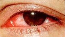
This is a frequent ambulatory complaint encountered in the ED. While many cases are secondary to benign conjunctiva irritation, the EP needs to be aware of vision threatening and systemic causes of �red eye� that demand immediate treatment and further work-up.
Conjunctivitis
- conjunctiva inflammation has numerous etiologies with classification based on cause: bacterial, viral, fungal, allergic or chemical.
- Symptoms vary depending upon the etiology. In most cases, visual acuity is unaffected. Suspect other etiologies in those patients who complain of vision loss.
Bacterial Conjunctivitis

- Etiology: Streptococcus pneumoniae and Staphylococcus aureus are the most common causative organisms. Gram-negative bacilli such as H. influenzae are also implicated. Consider Pseudomonas in patients who wear contact lenses. N. gonorrhoeae and Chlamydia are special concerns in neonates and patients at risk for sexually transmitted disease (see discussion below).
- Presentation: Patients complain of foreign body sensation, eye redness and discharge.
- Examination: conjunctiva injection and purulent eye discharge are noted. Chemosis and eyelid edema are sometimes present.
- Diagnosis is clinical. Patients with recurrent, persistent or severe disease should have swabs sent for Gram stain and culture.
- Treatment: Broad-spectrum topical antibiotic such as sulfacetamide, a flouroquinolone or bacitracin-polymyxin compound. Drops are used for 5-7 days. Do not patch or prescribe steroids. Follow-up with the ED or ophthalmology is encouraged. The patient should notice improvement in symptoms within 2-3 days; continued problems
indicate persistent disease or an allergic reaction to the prescribed medication.
Gonococcal Conjunctivitis
- Risk factors: Occurs in individuals after direct contact with others who have gonococcal cervicitis/urethritis and neonates within the first 5 days of life.
- Presentation: Patients have hyperacute onset of severe bilateral eye redness, eyelid swelling and copious discharge. Adults often give a history of risk factors for sexually transmitted disease.
- Examination: Significant for severe eyelid edema, chemosis and conjunctiva injection. Discharge is purulent and copious. Patients may have rapidly progressive corneal involvement including ulcerations and perforation; fluoroscein staining is indicated.
- Diagnosis is often clinical but Gram stain and culture of fluid are indicated.
- Treatment:
- Systemic antibiotics are necessary for gonococcal disease. Less severe cases are managed with IM ceftriaxone. Neonates and adult patients with severe disease and/or corneal involvement should be hospitalized for IVantibiotics. It is also recommended to treat with doxycycline or azithromycin for concomitant Chlamydia.
Treat all sexual partners as well as parents of infected infants.
- Topical antibiotics are necessary for those patients with corneal involvement.
- Saline irrigation four times a day is continued until eye discharge has resolved.
- Ophthalmology consultation should be obtained emergently.
- Children and adolescents with gonococcal conjunctivitis need evaluation for possible sexual assault.
Chlamydia Conjunctivitis
- Risk factors: Sexual contact with individuals who have Chlamydia cervicitis/urethritis. Greater than 50% of patients have concomitant genital infection. Neonates are also at risk, but disease occurs later than gonococcus at 5 days to 5 wk of life.
- Presentation: Symptoms are similar to viral conjunctivitis (see below). Patients may also give a history of recent genital symptoms consistent with cervicitis/urethritis.
- Neonates and infants may also have respiratory symptoms from concomitant Chlamydia pneumonia.
- Examination: Findings are similar to viral conjunctivitis although discharge is typically mucopurulent.
- Diagnosis is usually based upon history and eye examination. For definitive diagnosis, swabs are sent for flourescent antibody testing, Giemsa staining or culture.
- Treatment: Consists of systemic doxycycline or other appropriate antibiotic. Sexual contacts also need to be treated. Children and adolescents need evaluation for possible sexual assault.
Viral Conjunctivitis
- Etiology: Respiratory viruses, esp. adenovirus.
- Presentation: Patients have eye redness, itching and watery discharge. They often have a concurrent or recent upper respiratory tract infection.
- Examination: Findings include conjunctiva injection, tearing, conjunctiva follicles, eyelid edema, watery to mucoid discharge and preauricular lymphadenopathy.
- Treatment is symptomatic with artificial tears, cool compresses and topical antihistamines and vasoconstrictors. Topical antiviral medications are not effective.
- Viral conjunctivitis can spread rapidly from the affected to unaffected eye and to other individuals. Patients need to be excused from work and be instructed to avoid touching their eyes and sharing items with others.
- Epidemic keratoconjunctivitis (EKC): This is very contagious disease also caused by adenovirus. Common settings include dormitories and military housing. In contrast to typical viral disease, the patients will not have any systemic or respiratory symptoms. Both the conjunctiva and cornea may be involved. Treatment is symptomatic.
Patient contact with others should be limited. Symptoms sometimes last up to 4 wk.
Allergic Conjunctivitis
- Presentation: Symptoms include a characteristic intense bilateral itching and tearing.
Patients give a history of allergies, atopy and seasonal symptoms.
- Examination: The conjunctiva may have marked chemosis with either hyperemia or pallor. Discharge is watery.
- Treatment includes cool compresses, topical/oral antihistamines, topical vasoconstrictors and removal of the allergen if possible. Topical steroids are sometimes prescribed in conjunction with ophthalmology.
Pingueculum and Pterygium
- A pingueculum is a band of fibrous tissue occurring secondary to conjunctiva degeneration. It begins near the palpebral fissure and extends in the direction of, but does not involve, the cornea. A pterygium is a continuation of this process with the fibrous tissue extending onto the cornea. Both occur more frequently on the medial aspect of the eye.
- Risk factors: Chronic sun/dust exposure and irritation.
- Presentation: Most patients are asymptomatic. Rarely, the tissue becomes inflamed and causes eye redness and discomfort. Patients with large pterygia can also experience vision loss if the tissue extends into the visual axis.
- Examination: The primary finding is fibrous tissue extending across the conjunctiva from the palpebral area with/without corneal involvement. This tissue may be whitish to yellow or may be injected and erythematous if inflamed.
- Treatment: Asymptomatic lesions do not require treatment. Local inflammation is treated with topical vasoconstrictors and artificial tears. Elective surgical removal is indicated for persistent inflammation and large pterygia that interfere with vision or use of contact lenses.
Corneal Ulcer
- Etiology: Staphylococcus, Streptococcus, Pseudomonas, Herpes simplex and fungi.
- Risk factors: The primary cause is chronic use of soft contact lenses. Trauma secondary to organic material such as tree branches is a risk for fungal disease.
- Presentation: Pain, tearing, photophobia, FB sensation and eye redness.
- Examination: Ulcerations are visible as a whitish infiltrate of the cornea that will stain with fluoroscein. Other findings include conjunctiva injection, eyelid edema, chemosis and AC cells and flare.
- Treatment: Corneal ulcerations are an ophthalmologic emergency. Rigorous topical antibiotic therapy is necessary with agents that provide coverage of both Staphylococcus and Pseudomonas such as ciprofloxacin. Eye patching and steroids are contraindicated.
Herpes Simplex Keratitis
Etiology: Herpes simplex virus (HSV), either primary or reactivation. HSV causes many pathologic conditions of the eye including periorbital ulcerations, conjunctivitis, corneal ulcers and uveitis. Probably the most often-mentioned eye complication is keratitis.
HSV keratitis presents as mild eye pain, FB sensation, eye redness, tearing and photophobia. Patients may give a history of herpes infection or immunocompromise.
Examination: The classic finding is a dendritic pattern of fluoroscein uptake. Associated skin ulcerations are sometimes present. In contrast to zoster, these lesions are not in a dermatomal pattern and cross the midline.
Treatment: Topical antivirals such as trifluridine or vidarabine are necessary if there is corneal involvement. Milder disease such as conjunctivitis is treated with symptomatic measures. Oral antivirals are beneficial for patients with immunocompromise.
Steroids are recommended for certain HSV manifestations but should be administered only by the ophthalmologist. All patients with HSV keratitis must have an emergent ophthalmology evaluation.
Herpes Zoster Keratitis
- Etiology: Herpes zoster virus (HZV). Ocular and periocular disease is secondary to reactivation of the virus that is dormant in the trigeminal nerve.
- Presentation: The classic presentation is a painful rash on one side of the forehead. The rash is sometimes preceded by a prodrome of malaise and local pain. HZV also causes numerous ocular pathologies including conjunctivitis, keratitis, uveitis, scleritis and optic neuritis. Any patient who presents with the typical HZV rash involving the nasal tip needs to be evaluated for keratitis and other ocular manifestations.
- Examination: The rash of HZV is vesicular and located in a dermatomal pattern on the forehead. The rash does not cross the midline except in the rare bilateral case.
Ocular findings vary depending upon the extent of involvement. These patients often have punctate corneal lesions. Dendritic corneal lesions are mentioned in association with HZV keratitis but are actually mucous deposits that stain poorly with fluoroscein.
They are not epithelial defects and can be removed with a Q-tip.
- Treatment: HSV dermatitis is treated with systemic acyclovir or other antiviral agent.
Topical antivirals are not indicated. Severely ill patients require admission and intravenous medications. HZV ocular disease is often managed with steroids in conjunction with ophthalmology. Ancillary treatment should include analgesics.
Uveitis
- Inflammation of the uvea which includes the iris, ciliary body and choroid. Uveitis affects anterior structures, posterior structures or both. Clear determination of involvement is sometimes difficult.
- Etiology: Causes include trauma, recent eye surgery, autoimmune disease, infection, malignancy and sarcoidosis. Some cases are idiopathic.
- Presentation: Symptoms include deep, throbbing eye pain; photophobia; eye redness and tearing. There is often a history of symptoms related to other organ systems.
- Examination: Patients have mild anisocoria with the affected pupil having sluggish reaction to light. conjunctiva injection is pronounced especially at the limbus. SLE reveals the presence of cells and flare in the AC and possibly an associated hypopyon or layering of white blood cells in the AC. IOP varies. VA is usually normal. When present, vision deficits are mild. The patient with nontraumatic iritis also needs a
thorough systemic evaluation to identify signs of associated disease.
- Diagnosis is clinical although laboratories and other ancillary studies are often indicated depending upon patient presentation and history.
- Treatment: Uveitis is often managed with cycloplegics and steroids although this is not always the case. Additional treatment with systemic antibiotics, antivirals or immunosuppressants is sometimes necessary. Care should be coordinated with both ophthalmology and internal medicine.
Episcleritis
- Etiology: Inflammation of the episcleral vessels occurs most frequently in young women with the majority of cases being idiopathic. Other risk factors include collagen vascular disease, autoimmune disease, inflammatory bowel disease, gout and infection.
- Presentation: Patients complain of eye redness, tearing and dull pain. Vision is usually unaffected.
- Examination: Injection is typically located in segments or patches. The primary differential diagnoses are conjunctivitis and scleritis (see below). The episcleral vessels are large and arrayed in a radial fashion. They will react to topical phenylephrine and are mobile when lightly manipulated with a Q-tip applied to the surface of the eye.
- Treatment is symptomatic with artificial tears and systemic anti-inflammatory medications.
Scleritis
- Etiology: Inflammation of the sclera is often caused by underlying systemic disease, most commonly connective tissue disease, vasculitis and infection. Many cases are idiopathic.
- Presentation: Patients complain of severe, deep eye pain. They also report eye redness, tearing and photophobia. Vision is normal or mildly affected. Depending upon the etiology, other systemic symptoms are also reported.
- Examination: There is prominent eye redness, and the globe is tender to palpation.
Injection secondary to scleritis will not diminish with topical phenylephrine as opposed to conjunctiva and episcleral inflammation. In addition, inflamed scleral vessels will not move when a Q-tip is lightly applied to the surface of the eye.
- Patients with scleritis need emergent ophthalmology evaluation and further work-up to determine underlying etiology. Treatment varies depending upon the extent of involvement and includes systemic steroids, immunosuppressants and nonsteroidal anti-inflammatories.
|